A counterintuitive fact about carotid intima media thickness guidelines is that the test's clinical status changed substantially within only a few years. In 2010, the ACC/AHA considered carotid intima-media thickness reasonable for selected asymptomatic adults at intermediate risk. By 2013, the AHA/ACC position had shifted to routine use not providing benefit for first-event ASCVD risk assessment, as documented in this review of guideline history and technical standards.
That change doesn't mean CIMT is useless. It means the result depends on what is measured, how it is measured, and what clinical decision follows. For executives and professionals seeking proactive cardiovascular care in Delaware, southeastern Pennsylvania, or Maryland, the better question isn't just whether to get CIMT. It's whether the test will clarify risk that remains uncertain after blood pressure, lipids, family history, and established risk models have been assessed.
Table of Contents
- Why Carotid Intima Media Thickness Guidelines Matter Now
- How AHA, ACC, ESC, and Mannheim CIMT Recommendations Diverge
- How CIMT Is Actually Measured and Reported
- How to Interpret CIMT Results and Thresholds
- CIMT Compared With Carotid Plaque and Coronary Artery Calcium
- How CIMT Findings Change Risk Management and Follow-Up
- Common Misconceptions About CIMT Results
- Practical Checklist Before a CIMT Consultation
Why Carotid Intima Media Thickness Guidelines Matter Now
The test became controversial for a precise reason
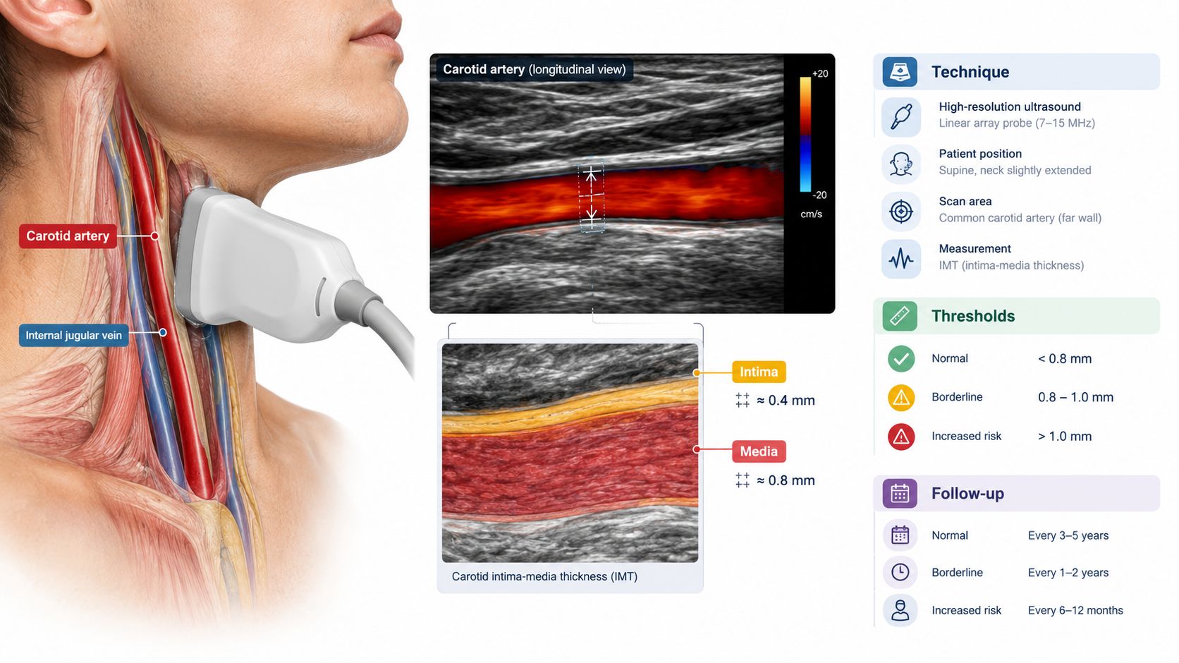
CIMT measures the thickness of the inner arterial layers, the intima and media, in the carotid artery. It can identify arterial wall remodeling before symptoms develop, but wall thickness isn't the same as focal plaque, and those findings don't carry identical clinical meaning.
Guideline interpretation changed because evidence and clinical priorities evolved. The 2010 ACC/AHA guidance described CIMT as reasonable for intermediate-risk asymptomatic adults, defined there as people with a 10% to 20% 10-year risk. The 2013 AHA/ACC update moved CIMT to Class III, meaning it shouldn't be used routinely for first ASCVD event risk assessment. That reversal over a short period is central to understanding why different clinicians still give different answers.
European guidance and the Mannheim consensus remained more open to selective carotid ultrasound, particularly when a patient has risk factors or sits in an intermediate-risk range. Their position emphasizes vascular hypertrophy, atherosclerosis detection, and technical consistency rather than treating one mean thickness value as a diagnosis.
Practical rule: A CIMT study earns its place when the result can change a decision. A measurement that produces no change in treatment, surveillance, or risk discussion adds little value.
The modern question is phenotype and protocol
A 50-year-old adult with intermediate estimated ASCVD risk, a strong family history, and borderline lipid results may reasonably want more information. In that setting, the clinically useful questions include:
- Is there focal carotid plaque, or only diffuse wall thickening?
- Was the measurement taken from a standardized, plaque-free far-wall segment?
- Was the result compared with an appropriate reference population?
- Would the finding change lipid treatment, blood pressure management, or the choice of another imaging study?
- Is there a credible plan for follow-up?
The evidence base increasingly supports interpreting plaque burden and measurement method more carefully than relying on one common-carotid mean thickness number. A systematic review summarized in the review of CIMT definitions and event associations found that combined-IMT and maximum common-carotid IMT were more strongly associated with events than some single-segment or mean definitions.
CIMT therefore remains a potential refinement tool, not a stand-alone verdict. Its value is greatest when the patient's risk is uncertain, the protocol is rigorous, and the result has a defined downstream action.
How AHA, ACC, ESC, and Mannheim CIMT Recommendations Diverge
The disagreement among major societies is real, but it's more specific than the phrase “CIMT is controversial” suggests. Some recommendations address routine risk prediction, while others address selective imaging and technical standards. Those are different questions.
Four positions, four emphases
| Society or group | Document or position | Role for CIMT | Role for carotid plaque | Practical implication |
|---|---|---|---|---|
| ACC/AHA | 2010 asymptomatic-adult guidance | Reasonable for selected intermediate-risk adults | Selective vascular assessment | CIMT could refine risk when the clinical decision remained uncertain |
| AHA/ACC | 2013 risk-assessment update | Routine first-event risk assessment not recommended | Plaque assessment receives greater practical attention | A mean CIMT result shouldn't automatically drive treatment |
| ESC and related European guidance | European prevention and vascular-risk guidance | More permissive selective use | Plaque can act as a risk modifier | Carotid imaging may help in selected patients with risk-factor uncertainty |
| Mannheim consensus | Carotid ultrasound consensus standards | Supports standardized IMT acquisition and reporting | Requires careful plaque-aware scanning | Technical quality is essential, regardless of whether CIMT is used for risk refinement |
The ACC summary of the earlier and later guideline positions describes the earlier permissive approach for intermediate-risk adults and the later movement away from routine CIMT use. Later summaries also report that major contemporary guidance has not recommended CIMT for systematic risk assessment because of limited standardization and limited incremental predictive value, as discussed in this guideline-focused review.
Where the recommendations converge
Despite their differences, the societies broadly agree on several principles:
- A standardized protocol matters. Measurements should be taken from the common carotid artery far wall, with careful attention to the bulb and plaque.
- No single cutoff works for everyone. Age, sex, ancestry, risk factors, and reference population influence interpretation.
- Plaque and diffuse wall thickness are not interchangeable. A focal lesion generally carries different information from a smooth, modest increase in wall thickness.
- CIMT shouldn't replace conventional risk assessment. Lipids, blood pressure, diabetes status, smoking, family history, and other established factors remain central.
The practical synthesis is straightforward. Plaque presence often drives the more consequential discussion, while mean CIMT may provide modest additional context when coronary artery calcium is unavailable, unsuitable, or inconclusive. A clinician should explain that distinction before ordering the study.
How CIMT Is Actually Measured and Reported
A credible CIMT report should allow another qualified reader to understand exactly what was measured. A number without an acquisition protocol, anatomical location, or reference framework is difficult to interpret.
The core acquisition protocol
CIMT is generally obtained with B-mode ultrasound, using a high-frequency linear probe. Peer-reviewed reviews commonly describe a probe frequency of at least 7 MHz, while many clinical protocols use a higher frequency when image quality permits. The examination should focus on the distal common carotid artery, ideally on a straight, plaque-free segment at least 5 mm below the carotid bulb.
The far wall is preferred because its interfaces can be identified more consistently than the near wall. The sonographer looks for the lumen-intima boundary and the media-adventitia boundary, then measures the tissue between them. ECG gating can help reduce variability related to the cardiac cycle, and automated edge-detection software may improve consistency when the interfaces are clearly visible.
The technical standards for reproducible CIMT acquisition emphasize the far wall of the distal common carotid artery, plaque-free straight segments, appropriate imaging equipment, and segmentation-specific reporting.
What should appear in the report
A useful report should identify:
- The side measured, including right and left carotid arteries.
- The arterial segment, such as the distal common carotid far wall.
- The measurement approach, including whether acquisition was B-mode and ECG-gated.
- Mean and maximum values, when both are clinically appropriate.
- The reference framework, such as age-, sex-, or population-based percentile interpretation.
- Plaque findings, including whether focal lesions were present, absent, or inadequately assessed.
- Image quality and reader credentials, particularly when the result will influence treatment.
Common weaknesses include low-frequency imaging, oblique probe angles, near-wall-only sampling, inconsistent measurement locations, and a report that gives CIMT but doesn't discuss plaque. A study that doesn't document its protocol should be treated cautiously, even when the numerical result appears precise.
How to Interpret CIMT Results and Thresholds
CIMT interpretation relies on two broad frameworks. The first uses absolute thresholds. The second compares the result with a reference population using percentiles. Neither approach creates a universal diagnosis.
Thresholds are reference points, not verdicts
European guidance has often treated CIMT of 1.0 mm or more as abnormal, while some expert sources use values above 0.9 mm in selected settings. The ASE framework considers CIMT at or above the 75th percentile of a healthy reference population to represent higher risk. The 25th to 75th percentile range is generally considered average and doesn't automatically alter traditional risk estimates, as summarized in this review of CIMT thresholds and interpretation frameworks30117-0/fulltext).
Those values should not be presented as universal disease cutoffs. A result can be above a threshold without proving obstructive disease, and a result below a threshold doesn't eliminate risk from LDL-related exposure, hypertension, diabetes, smoking, lipoprotein(a), family history, or plaque elsewhere.
| Age band | Typical mean CIMT | 75th percentile | Clinical interpretation |
|---|---|---|---|
| Adult reference populations | Varies by population | Must be population-specific | Use the laboratory's validated reference set |
| Younger adults | Often lower than older adult values | Requires age- and sex-matched comparison | A modest absolute value may still be high for age |
| Middle-aged adults | Increases across populations with age and risk factors | Not universal | Combine percentile, plaque status, and ASCVD risk |
| Older adults | Greater biological variability | Reference population becomes especially important | Avoid interpreting age-related thickening as a diagnosis |
Read the whole report, not one line
A careful interpretation asks whether the value is:
- Mean or maximum, because maximum measurements can capture a focal area that a mean may dilute.
- Right or left, since the sides may differ.
- Far-wall distal common carotid, or another segment with different reference expectations.
- Plaque-free, because plaque can distort the meaning of nearby wall measurements.
- Compared with an appropriate population, rather than a generic normal range.
A practical resource on minimum diagnostic standards for 2026 can help readers evaluate whether an advanced diagnostic report contains enough technical information to support a meaningful clinical discussion.
Why women and younger adults need context
There isn't a widely accepted universal CIMT cutoff that works equally well for women, younger adults, and people from different ancestry backgrounds. Genetic architecture and vascular risk patterns can differ across ancestries, which makes one-size-fits-all interpretation unreliable.
CIMT is best understood as a continuous risk marker. The result should be placed alongside plaque burden, LDL-related markers, blood pressure, diabetes status, family history, and an established risk model. The most useful clinical question is rarely “Is this normal?” It's “Does this finding materially change the person's prevention plan?”
CIMT Compared With Carotid Plaque and Coronary Artery Calcium
CIMT, carotid plaque, and coronary artery calcium answer related but different questions.
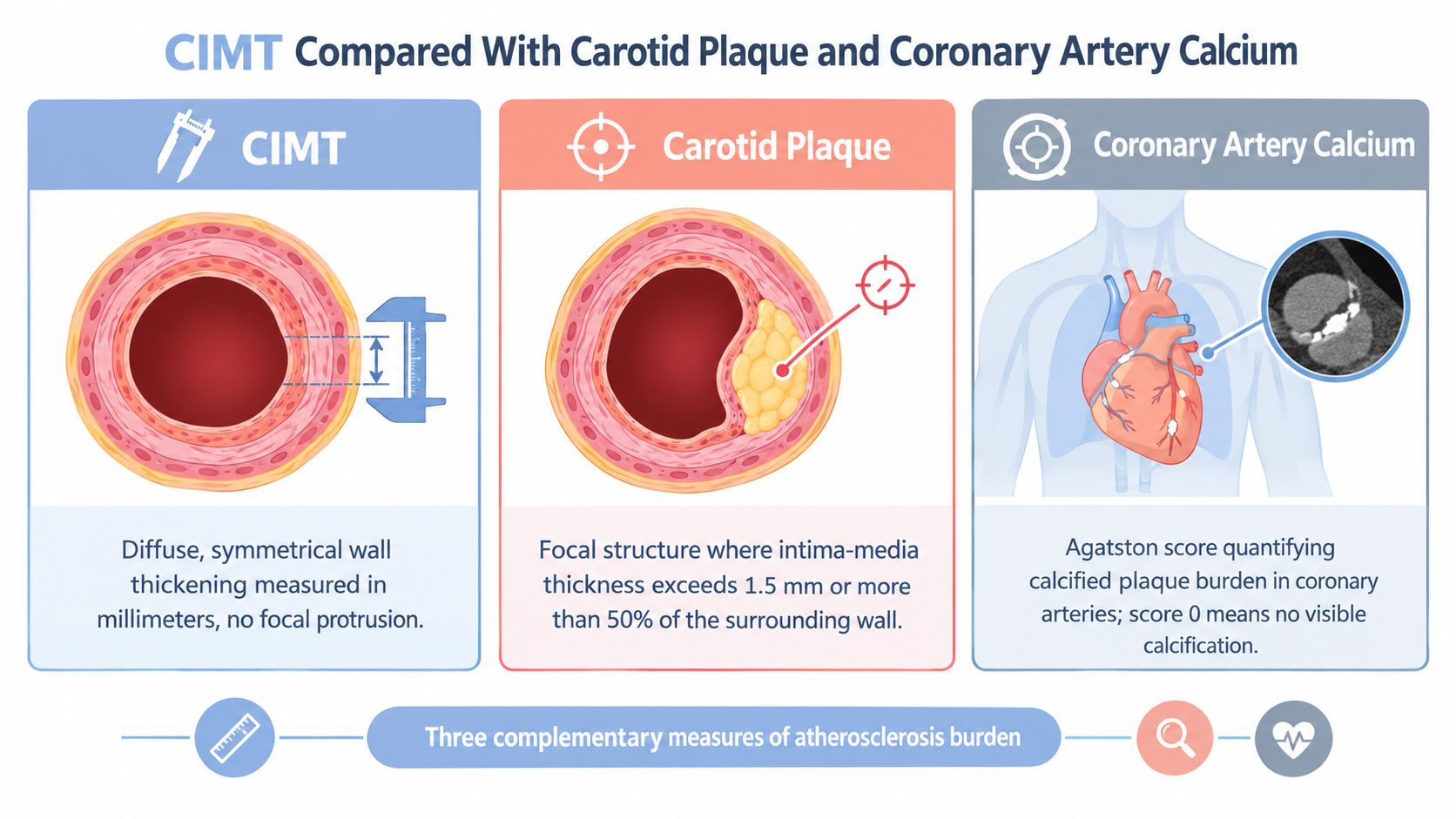
CIMT measures diffuse wall thickness. Carotid plaque identifies a focal arterial wall abnormality. Coronary artery calcium, obtained with non-contrast CT, quantifies calcified plaque in the coronary arteries using an Agatston score. CIMT is radiation-free but operator-dependent. CAC involves radiation exposure but offers a more direct assessment of calcified coronary atherosclerotic burden.
Current practice generally gives greater decision weight to plaque detection and CAC than to mean CIMT alone. Carotid plaque can demonstrate focal atherosclerosis, while CAC can help resolve uncertainty about statin treatment in selected adults. CIMT may still be complementary, particularly when a younger or intermediate-risk patient needs additional vascular context and CAC isn't the preferred first study.
Choosing the modality depends on the decision
| Measure | What it reveals | Main strengths | Main limitations |
|---|---|---|---|
| CIMT | Diffuse arterial wall thickness | Noninvasive, no radiation, accessible | Operator-dependent and sensitive to protocol |
| Carotid plaque | Focal arterial wall disease | Directly identifies focal atherosclerosis | Requires careful plaque imaging and reporting |
| CAC | Calcified coronary plaque burden | Standardized coronary calcium quantification | Doesn't show non-calcified plaque and uses CT |
A patient seeking a broader overview of available studies can review this guide to advanced cardiovascular imaging. The decision should remain clinical, not consumer-driven. A test is useful when the result changes the conversation about treatment, intensity, or follow-up.
How CIMT Findings Change Risk Management and Follow-Up
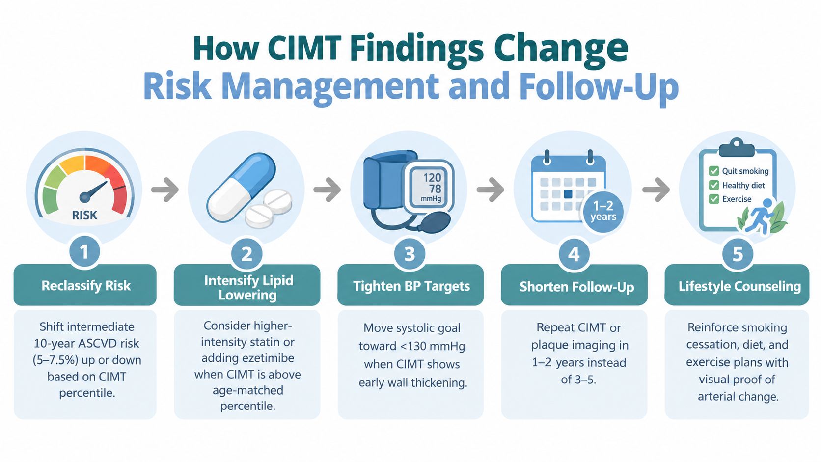
CIMT should influence the pace and intensity of prevention, not act as an isolated prescription generator. A higher percentile or a plaque finding may strengthen the case for addressing modifiable risk factors, but the response still depends on the full ASCVD profile.
Connect the result to a specific action
A clinician may use the result to revisit:
- Risk classification, especially when the estimated 10-year risk is intermediate or the history suggests risk may be underestimated.
- Lipid management, including whether the intensity of LDL-lowering therapy matches the overall risk profile.
- Blood pressure control, with the treatment target individualized to the patient's health, tolerance, and comorbidities.
- Lipoprotein(a) and ApoB testing, when inherited or particle-related risk remains unclear.
- Lifestyle structure, including nutrition, exercise, sleep, tobacco avoidance, and weight management.
- Additional imaging, particularly when carotid findings and the clinical picture don't align.
A normal CIMT without plaque may provide reassurance in a low-to-intermediate-risk patient, but it doesn't justify ignoring persistent risk factors. Conversely, higher CIMT or plaque commonly prompts more deliberate prevention and a clearer follow-up plan rather than urgent invasive testing.
Follow-up should be purposeful
Repeat imaging shouldn't be scheduled just because a patient wants a new number. The interval depends on baseline risk, plaque status, treatment changes, image quality, and whether a future result would alter management. Serial measurements are most useful when the same or a comparable protocol is used and the clinician can explain what constitutes a meaningful change.
Patients who want to find their heart disease risk can use a risk calculator as an educational starting point, but a calculator can't replace clinical interpretation. ApoB may also clarify the number of atherogenic particles, as discussed in this guide to the ApoB test.
Antiplatelet therapy deserves particular caution. CIMT alone shouldn't trigger aspirin or another antiplatelet medication. Such treatment depends on the balance between cardiovascular benefit and bleeding risk, along with the patient's broader clinical phenotype.
Common Misconceptions About CIMT Results
A normal result doesn't erase risk
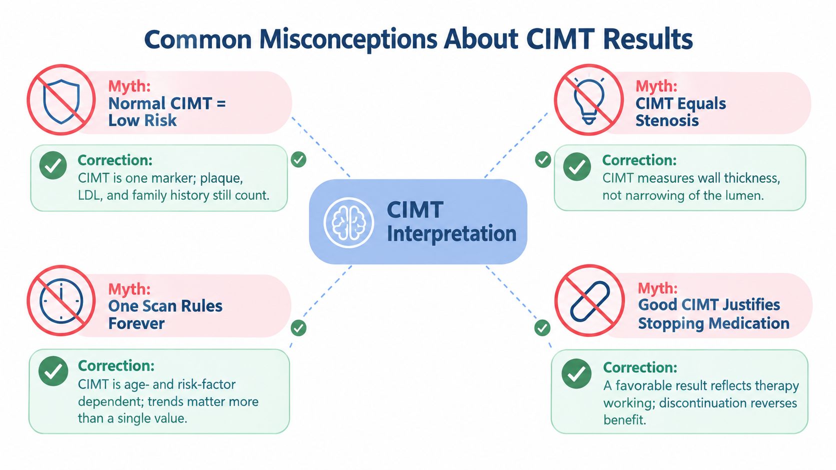
A normal CIMT result means that the measured arterial wall doesn't show an abnormality under that protocol and reference framework. It doesn't mean the coronary arteries are free of disease, and it doesn't offset raised LDL exposure, high blood pressure, diabetes, smoking, family history, or inherited lipoprotein risk.
A favorable result can be useful. It may support a measured approach when treatment decisions are uncertain, but it shouldn't override stronger evidence from other risk domains.
An elevated result isn't automatically a diagnosis
CIMT measures wall thickness, not stenosis and not necessarily a clinically significant obstructive lesion. An increased value can signal increased vascular risk or arterial remodeling, but the report must be interpreted with plaque status, image quality, and the patient's age and risk profile.
The reverse misconception is also common. A patient may believe that a high number demands urgent invasive workup. In most asymptomatic settings, the more immediate consequence is a discussion about prevention, not emergency intervention.
One scan doesn't define a lifetime trajectory
Vascular measurements vary with technique, sonographer skill, arterial segment, blood pressure, and biological change. A single scan offers a snapshot, not a guarantee about future progression or regression. Serial testing may help in selected cases, but it should occur on clinically sensible intervals rather than repeated frequently without a decision attached.
The right interpretation is calibration, not catastrophe. CIMT helps place prevention in context. It doesn't provide permission to stop treatment, and it doesn't prove that an urgent procedure is needed.
Medication decisions remain broader than CIMT
A favorable scan may reflect effective prevention, favorable biology, or limitations of the measurement. It shouldn't be used on its own to discontinue a medication that has a well-established indication. Similarly, an unfavorable scan should prompt a structured review of lipids, pressure, glycemia, tobacco exposure, exercise, nutrition, sleep, and adherence before treatment is intensified.
Practical Checklist Before a CIMT Consultation
A productive consultation begins before the ultrasound. The clinician needs enough information to determine whether CIMT can answer a meaningful question and how the result would fit into the prevention plan.
Bring the clinical context
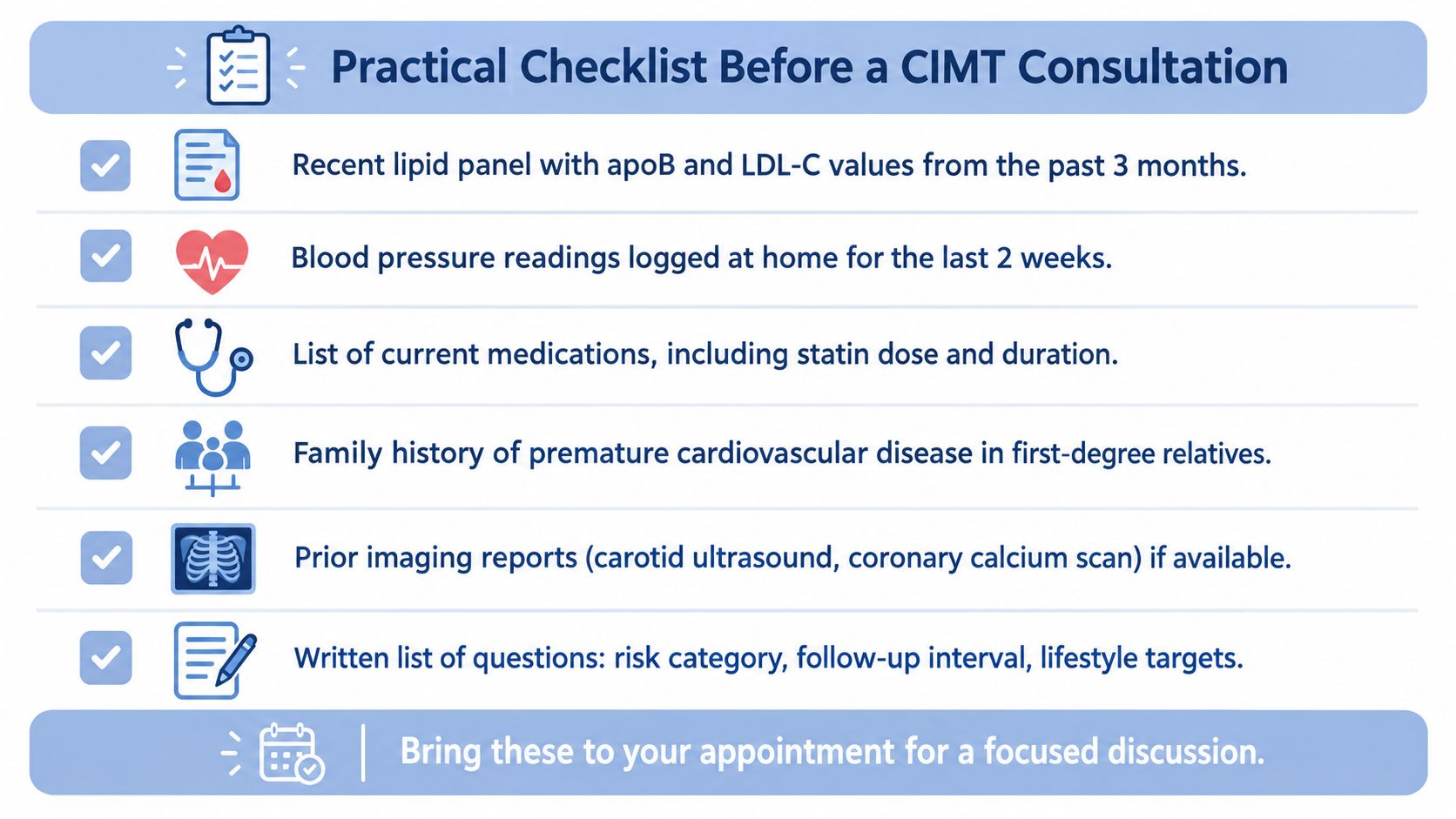
Patients should gather:
- Recent lipid data, including LDL-C and ApoB when available.
- Blood pressure information, including home readings if office values vary.
- Family history, especially premature ASCVD in first-degree relatives.
- Medication details, including names, doses, duration, and tolerance.
- Lifestyle factors, such as smoking status, exercise patterns, sleep, nutrition, and work-related travel.
- Prior imaging, including carotid ultrasound or coronary calcium results.
The goal isn't to arrive with a perfect dossier. It's to help the physician distinguish a measurement that adds useful information from one that produces another isolated number.
Ask about the protocol
Before scheduling the study, practical questions include:
- Will the examination assess carotid plaque as well as CIMT?
- Will measurements come from the far wall of the distal common carotid artery?
- Is the acquisition performed with B-mode ultrasound and appropriate high-frequency equipment?
- Will the report include right and left sides, mean and maximum measurements where relevant?
- Is automated edge detection used, and will a qualified physician review the images?
- Will the result be compared with an appropriate age-, sex-, and population-specific reference?
- What action would follow a higher percentile, increased thickness, or plaque finding?
- What finding would justify another modality, such as CAC?
- When would repeat imaging provide clinically useful information?
A report should specify the arterial segment, wall location, measurement method, image quality, reference framework, and plaque assessment. If those elements are absent, the number may be less informative than it appears.
A printable summary card
Before the appointment
- Bring lipid, blood pressure, medication, family history, and prior imaging information.
- Ask whether plaque imaging is included.
- Confirm far-wall common-carotid measurement and bilateral reporting.
- Ask how the result will alter ASCVD risk assessment.
- Clarify whether lipid or blood pressure treatment should change.
- Agree on a follow-up interval based on a specific clinical purpose.
For adults comparing physician-led options in the region, this overview of a preventive cardiologist near you can help frame questions about diagnostic scope and clinical interpretation. Testing is available to anyone, not only members of an executive health program, but every study should be tied to a clear medical question and reviewed with a qualified clinician.
Hosmane Longevity Center offers physician-led preventive cardiology, advanced cardiac testing, and clinical interpretation of CIMT, carotid plaque, CAC, and related biomarkers for adults seeking a clearer view of subclinical cardiovascular risk. Visit Hosmane Longevity Center to request a consultation in Lewes or Wilmington and discuss which testing approach fits the individual risk profile.