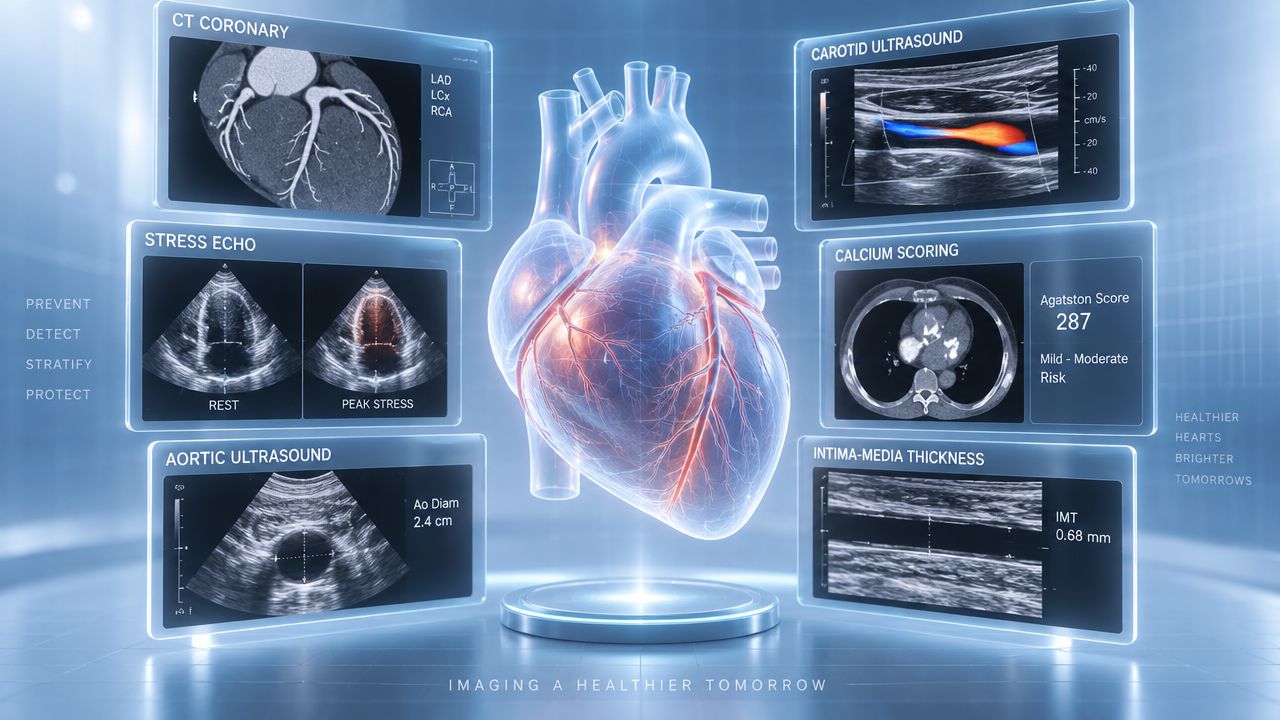
Advanced cardiovascular imaging isn't a universal screening package, and more testing isn't automatically better care. Each modality answers a different clinical question: How much coronary plaque is present? Is there vascular disease? Does exertion reveal ischemia? Is an artery narrowed, or is the heart structurally abnormal? The useful test is the one whose result could change a clearly defined decision.
That distinction matters for executives and professionals in Lewes, Wilmington, Rehoboth, and nearby Pennsylvania and Maryland, where preventive care often has to fit around demanding schedules and complex medical histories. Adults may discuss focused testing or an executive health program with Dr. Vinay Hosmane at Hosmane Longevity Center, where testing is available to anyone when clinically appropriate, not only to program members.
The evidence also argues against blanket screening. Current guidance supports selective use of imaging when symptoms, baseline risk, family history, biomarkers, or uncertainty around treatment make additional information useful. Results require clinician interpretation, because a scan can identify risk without explaining the best next step on its own. The following eight modalities are organized by the questions they answer, from silent plaque burden to integrated prevention.
Table of Contents
- 1. Coronary Artery Calcium Scoring
- 2. Carotid Intima-Media Thickness Ultrasound
- 3. Stress Echocardiography
- 4. Advanced Carotid Doppler Ultrasound With Flow Assessment
- 5. Ankle-Brachial Index With Exercise Testing
- 6. Abdominal Aortic Ultrasound Screening
- 7. High-Resolution Cardiac CT
- 8. Core Cardiovascular Assessment at Hosmane Longevity Center
- 8-Modality Advanced Cardiovascular Imaging Comparison
- Turn Images Into a Prevention Plan
1. Coronary Artery Calcium Scoring
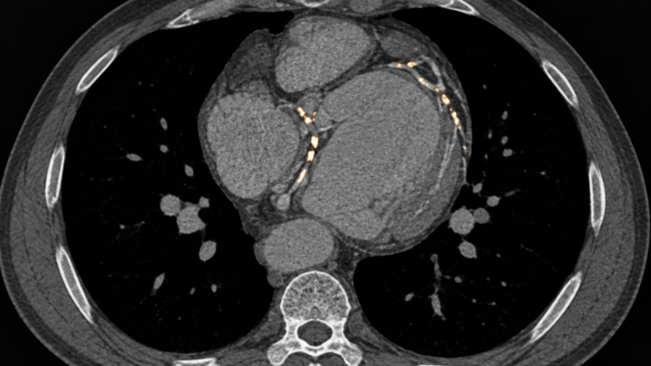
Coronary artery calcium, or CAC, answers a focused question: how much calcified plaque is present in the coronary arteries, and could that finding change prevention? A noncontrast CT scan detects calcium within atherosclerotic plaque. It cannot show every form of plaque, but it can identify coronary atherosclerosis before symptoms appear.
The 2019 ACC/AHA primary-prevention guideline assigns CAC a selective role for adults at intermediate risk when standard risk assessment leaves uncertainty about statin therapy. A CAC score of 0 can support deferring a statin in selected patients. CAC ≥100 Agatston units or at least the 75th percentile supports statin initiation in the appropriate clinical context. The global CAC guidance summary likewise presents CAC staging as a tool for setting the intensity of lipid lowering and risk-factor management, not as a simple normal-or-abnormal label.
Practical rule: CAC has the clearest value when its result can resolve a treatment decision. It is not a universal screening test for people at very low risk or for patients with established coronary disease.
A 52-year-old executive with a strong family history and uncertainty about medication might discuss CAC with a preventive cardiologist. A high result could support more intensive prevention. A zero result might reduce uncertainty for a carefully selected patient. Neither finding replaces attention to blood pressure, lipids, exercise, nutrition, sleep, and other modifiable risks.
The 2019 ACC/AHA framework also suggested considering repeat CAC after 5 years in selected adults with scores of 0 or 1–99. That interval is not an automatic appointment. Changing risk factors and the likelihood that another scan would alter management should guide the decision.
2. Carotid Intima-Media Thickness Ultrasound
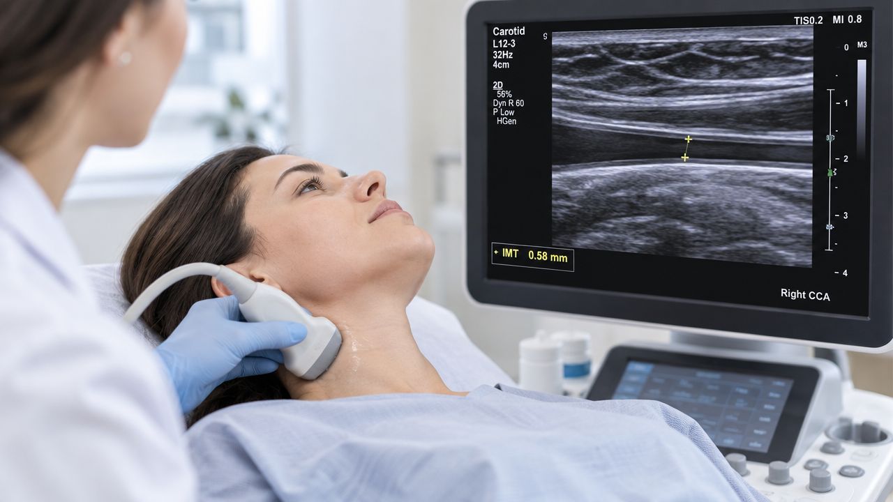
Carotid intima-media thickness, or CIMT, measures the thickness of layers in the wall of the carotid artery using ultrasound. It sounds straightforward, but its value depends heavily on image quality, measurement technique, and the clinical question. A thicker arterial wall can reflect vascular remodeling and atherosclerotic burden, yet that finding doesn't carry the same decision clarity as visible carotid plaque or coronary calcium in many patients.
The ACC/AHA risk-assessment review states that CIMT shouldn't be used routinely for first-event ASCVD risk prediction because evidence and standardization have been insufficient. A newer review of vascular imaging describes CIMT as a selective adjunct, particularly in intermediate-risk situations, while noting that carotid plaque and CAC often compete as stronger or more actionable markers.
That creates an important distinction for a professional who already has advanced lipid testing. CIMT may add context about arterial structure, but it shouldn't automatically be ordered because it is noninvasive. If the result won't change lipid treatment, blood-pressure management, or the need for another study, its practical value may be limited.
Where CIMT can still fit
CIMT can support a broader vascular discussion when family history or overall risk appears difficult to classify. The examination is painless and uses no radiation, but its usefulness relies on a cardiovascular sonographer and interpreting clinician who follow consistent acquisition and reporting methods. A report should distinguish diffuse wall thickening from focal plaque, because those findings don't carry identical implications.
A patient in Wilmington with high ApoB, a family history of early disease, and otherwise borderline risk might discuss whether CIMT adds meaningful information after the lipid evaluation. Another patient with clearly high risk may need treatment regardless of CIMT, making the scan less likely to change care.
For readers exploring how preventive testing can be integrated with broader cardiovascular planning, the Hosmane Longevity Center preventive cardiology resources provide additional context. The appropriate role of CIMT remains selective, not automatic.
3. Stress Echocardiography
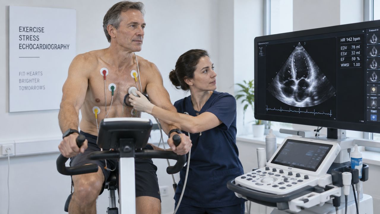
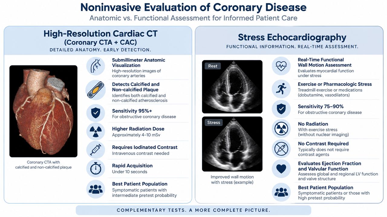
Stress echocardiography answers a different question from CAC. Instead of estimating accumulated plaque burden, it asks whether the heart develops evidence of inducible ischemia when its workload rises. Ultrasound images are obtained at rest and during exercise or medication-induced stress, allowing the clinician to assess changes in regional wall motion, pumping function, valve behavior, and hemodynamic response.
This makes stress echocardiography more relevant to a patient with exertional chest pressure, unexplained shortness of breath, known coronary disease, or an abnormal preliminary evaluation than to an asymptomatic person seeking a broad screening package. Exercise is generally preferable when a patient can perform it because the test also reveals exercise capacity, blood-pressure response, symptoms, and electrocardiographic changes. Pharmacologic stress can be useful when exercise isn't feasible or when the clinical question requires another approach.
At Hosmane Longevity Center, this modality is positioned as a focused study rather than an automatic component of every preventive evaluation. That reflects the broader evidence-based principle that imaging should be selected for clinical utility, not because a technology is available.
A normal stress study can be reassuring about inducible ischemia, but it doesn't prove that coronary plaque is absent.
Consider a professional with exertional discomfort and several cardiovascular risk factors. A stress echocardiogram that shows a convincing abnormal wall-motion response may lead to an anatomic study or specialist coordination. A normal result may shift attention toward other causes of symptoms, such as pulmonary disease, rhythm disturbance, valvular disease, or diastolic dysfunction. The result must be interpreted alongside symptoms, ECG findings, biomarkers, and prior imaging.
Stress echocardiography also illustrates why “more advanced” doesn't always mean “better.” CAC can clarify long-term prevention in selected asymptomatic adults, while stress echocardiography evaluates a functional question. Ordering the wrong test can produce information that is accurate but not useful for the decision at hand.
4. Advanced Carotid Doppler Ultrasound With Flow Assessment
Carotid Doppler answers an anatomic and hemodynamic question: Is plaque narrowing a carotid artery enough to alter blood flow, and does the finding fit the patient's neurologic history? B-mode ultrasound shows arterial structure and plaque, while Doppler evaluates blood-flow velocity through the vessel. The combined examination can help identify suspected stenosis and guide the need for confirmatory imaging.
This test has a different place in care from CIMT. CIMT describes arterial wall thickness. Doppler focuses on flow and narrowing. The distinction becomes especially important after transient neurologic symptoms, such as transient monocular vision loss or a transient ischemic attack, when identifying clinically important carotid disease may affect urgent specialist evaluation.
A patient with a recent TIA and a concerning Doppler result may need CT or MR angiography before an intervention is considered. Ultrasound findings can differ from cross-sectional imaging, and treatment decisions depend on symptoms, degree of stenosis, plaque features, overall health, and specialist assessment. An asymptomatic finding shouldn't be interpreted the same way as a similar narrowing in someone with a recent neurologic event.
What the scan can and cannot establish
Doppler can show abnormal velocity and disturbed flow, but velocity alone doesn't determine stroke risk. Plaque morphology, symptom timing, blood pressure, smoking status, lipid exposure, and the presence of disease elsewhere all matter. Vertebral artery flow can also add information, particularly when clinicians are evaluating unusual flow patterns or possible subclavian disease.
For a 60-year-old executive with no neurologic symptoms, broad carotid screening may provide less actionable information than targeted prevention based on blood pressure, lipids, and coronary risk. For an older adult with a TIA history or known carotid plaque, the test may answer a direct surveillance or referral question.
That is why a normal carotid Doppler doesn't erase coronary risk, and a high CAC score doesn't automatically imply carotid stenosis. The arterial system is connected, but each modality examines a different part of the disease process.
5. Ankle-Brachial Index With Exercise Testing
The ankle-brachial index, or ABI, asks whether leg arteries deliver blood adequately at rest. Clinicians compare systolic pressure at the ankle with pressure in the arm. If symptoms occur only during walking, exercise ABI can reveal a perfusion problem that a resting measurement misses.
The American Academy of Family Physicians review of ABI testing identifies ≤0.90 as an abnormal ABI threshold and explains why exercise testing can uncover peripheral arterial disease when resting values appear normal. This makes ABI relevant to people with exertional calf discomfort, diminished pulses, unexplained walking limitation, or atherosclerotic risk that isn't fully explained by coronary testing.
A 62-year-old executive who stops walking because of calf discomfort may have a normal resting ABI but an abnormal post-exercise result. That finding doesn't establish the complete anatomy, but it can justify duplex ultrasound and a broader evaluation of peripheral arterial disease. A different patient with leg fatigue may have a normal exercise ABI, directing attention toward orthopedic, neurologic, medication-related, or conditioning causes.
Read ABI in context
ABI isn't a standalone measure of whole-body cardiovascular health. It should be considered alongside symptoms, pulses, blood pressure in both arms, smoking history, diabetes, lipid markers, and evidence of disease in other arterial beds. An abnormal result may change the intensity of preventive treatment, but it doesn't by itself identify which artery is narrowed or whether an intervention is needed.
The test also has limitations. Arterial calcification can make ankle pressures appear artificially high, so a suspicious clinical picture with a noninterpretable ABI may require another physiologic assessment, such as toe pressure or pulse-volume recording. A large difference between arm pressures can also prompt evaluation for upper-extremity arterial disease.
ABI therefore offers a useful counterpoint to coronary imaging. CAC estimates coronary plaque burden, while ABI detects impaired lower-extremity perfusion. Neither replaces the other, and neither should be ordered to make an assessment look complete.
6. Abdominal Aortic Ultrasound Screening
Abdominal aortic ultrasound answers a focused structural question: Is the abdominal aorta enlarged in a way that requires surveillance or vascular consultation? The examination uses ultrasound to measure aortic diameter and can identify an abdominal aortic aneurysm without radiation or contrast.
The strongest screening rationale applies to selected older adults, especially men with a smoking history. For other groups, including women and younger adults, the decision is more individualized because the balance between detection, surveillance, incidental findings, and downstream care differs. A preventive visit should therefore identify the risk profile before adding abdominal imaging.
A former smoker with hypertension may reasonably discuss abdominal aortic ultrasound as part of a vascular evaluation. If enlargement is found, the result usually creates a surveillance question rather than an immediate treatment decision. A normal scan can close that particular screening question, but it doesn't assess coronary plaque, carotid disease, or leg perfusion.
Why incidental findings matter
Ultrasound can reveal findings outside the aorta, including abnormalities involving nearby abdominal organs. Those findings may be benign, but they still require documentation and communication with the primary clinician when follow-up is appropriate. The value of screening includes not only detecting an aneurysm, but also managing what the scan reveals without creating confusion.
A patient in Rehoboth who requests “every available scan” may not benefit from abdominal aortic imaging if the person lacks a relevant risk profile. Conversely, an older adult with a meaningful smoking history may have a clear clinical question that makes the test sensible. The decision should be tied to whether the result would establish surveillance, specialist referral, or reassurance about aneurysm risk.
Abdominal aortic ultrasound also demonstrates the importance of scope. A technically simple test can be appropriate in one risk context and low-value in another. Preventive cardiology works best when each image has a defined purpose and a responsible follow-up plan.
7. High-Resolution Cardiac CT
High-resolution cardiac CT answers two different clinical questions. CAC scoring uses noncontrast CT to measure calcified coronary plaque and refine prevention decisions in selected asymptomatic adults. Coronary CT angiography, or CCTA, uses contrast to show the coronary lumen and plaque, usually when symptoms or prior findings raise a question about coronary anatomy.
CCTA is not a more detailed CAC scan. CAC addresses estimated plaque burden and treatment uncertainty. CCTA examines anatomy in a patient with a defined diagnostic question. The ACC/AHA primary-prevention guideline supports selective CAC use, while Hosmane Longevity Center testing information describes the center's broader approach to cardiovascular diagnostics.
A patient with atypical chest discomfort and an intermediate clinical probability of coronary disease may discuss CCTA with a cardiologist. A clear study can help exclude important obstructive anatomy. An equivocal result, or extensive calcification that limits interpretation, may lead to functional testing or another evaluation. Contrast exposure, kidney function, heart rate, rhythm, and possible downstream procedures should be considered before testing.
Anatomy isn't the same as physiology
CCTA shows whether an artery appears narrowed, but an anatomic narrowing does not always limit blood flow during exertion. Symptoms, ECG findings, biomarkers, and stress imaging may therefore remain relevant before invasive evaluation or revascularization is considered.
CCTA has less value as a screening test for an asymptomatic person without a defined clinical question. Borderline or incidental findings can prompt additional testing without changing prevention or treatment. For someone with symptoms and an uncertain diagnosis, that trade-off may be reasonable. For a low-risk asymptomatic adult, the added information may not justify contrast exposure, interpretation limits, and follow-up.
The practical distinction is straightforward: risk refinement and symptom evaluation require different tests. CAC estimates calcified plaque burden. CCTA examines coronary anatomy. Related technology does not make the studies interchangeable, and more imaging is useful only when the result can change the next decision.
8. Core Cardiovascular Assessment at Hosmane Longevity Center
The Core Cardiovascular Assessment answers the broadest question: Which combination of findings can clarify cardiovascular risk, identify a meaningful abnormality, and create a prioritized prevention plan? It isn't a single image. It is an integrated evaluation that can combine selected imaging, physiologic testing, laboratory markers, risk modeling, and physician interpretation.
The value of an integrated assessment lies in connecting results that are often separated across appointments. CAC may speak to coronary plaque burden. ABI may identify peripheral arterial disease. Carotid imaging may add information about vascular structure or flow. Stress echocardiography may address ischemia or functional reserve when symptoms justify it. Advanced lipid markers such as ApoB and Lp(a), blood pressure monitoring, ECG findings, sleep evaluation, exercise capacity, and nutrition history can help determine what the images mean in context.
The ACC/AHA CAC analysis illustrates why guideline framework matters. CAC eligibility varied substantially across ACC/AHA Pooled Cohort Equation, ACC/AHA PREVENT, and ESC/EAS approaches, so the same adult may be considered for additional risk refinement under one framework but not another. That isn't a contradiction. It reflects differences in how guidelines define uncertainty and treatment thresholds.
Integration should reduce unnecessary testing
The outcomes-based cardiovascular imaging review describes a mature imaging field that evaluates clinical outcomes, safety, cost, and quality of life, not only image quality. That standard supports a restrained approach. A good assessment doesn't accumulate abnormalities. It identifies which findings deserve action and which don't.
At Hosmane Longevity Center, the executive health program is designed around physician-led preventive evaluation, with in-person care in Delaware and cardiovascular-specific diagnostics interpreted in clinical context. Testing is available to anyone when appropriate, whether or not the individual enrolls in an executive health program.
A Wilmington professional with a strong family history, high Lp(a), and an unclear statin decision may need a different evaluation from a Lewes adult with exertional symptoms. The integrated model is useful only when the testing sequence follows the clinical question and produces a plan that fits the person's schedule, risks, and preferences.
8-Modality Advanced Cardiovascular Imaging Comparison
| Item | Implementation complexity | Resource requirements | Expected outcomes | Ideal use cases | Key advantages |
|---|---|---|---|---|---|
| Coronary Artery Calcium (CAC) Scoring | Low, quick non-contrast CT | CT scanner, low radiation (~1 mSv), minimal staff, <10 min | Quantitative Agatston calcium score; coronary atherosclerotic burden and 5–10 yr risk stratification | Asymptomatic adults 40–75 with intermediate risk to guide preventive therapy | High specificity; zero score strong negative predictive value; standardized and reproducible |
| Carotid Intima-Media Thickness (CIMT) Ultrasound | Moderate, operator-dependent ultrasound | High‑resolution ultrasound, certified sonographer, 15–20 min | IMT measurement and focal plaque detection; incremental stroke/MI risk estimate | Intermediate‑risk individuals or those with family history of early CVD | Radiation‑free, inexpensive, suitable for serial monitoring; visualizes vessel wall structure |
| Stress Echocardiography (Exercise or Pharmacologic) | High, stress protocol plus imaging | Echo machine, exercise equipment or pharmacologic agents, monitoring staff, 30–60 min | Detection of reversible ischemia via wall‑motion abnormalities; functional cardiac assessment | Symptomatic patients or those with abnormal screening tests needing ischemia evaluation | High diagnostic accuracy for obstructive CAD; assesses function, valves, and prognosis; no radiation with exercise |
| Advanced Carotid Doppler Ultrasound with Flow Velocity Assessment | Moderate–High, B‑mode + Doppler, technical interpretation | Doppler ultrasound, experienced sonographer, optional contrast, 20–30 min | PSV/EDV and plaque morphology; grades hemodynamic stenosis and identifies high‑risk plaques | Symptomatic cerebrovascular patients, surveillance of known carotid disease, older adults | Accurate for hemodynamically significant stenosis; identifies ulceration/neovascularization non‑invasively |
| Ankle‑Brachial Index (ABI) with Exercise Testing | Low, simple bedside protocol | Portable Doppler, cuffs, treadmill for exercise ABI, ~10–20 min | Resting and exercise ABI ratios; detects PAD and exercise‑induced perfusion deficits | Screening for PAD in at‑risk or older adults; evaluation of exertional leg symptoms | Inexpensive, portable, rapid; strong predictor of CV mortality and identifies occult PAD |
| Abdominal Aortic Ultrasound Screening (AAA) | Low, focused abdominal ultrasound | Ultrasound machine, trained operator, 10–15 min | Aortic diameter measurement; detection and surveillance of AAA (≥3.0 cm) | Men 65–75 with smoking history and other high‑risk older adults | Excellent sensitivity/specificity for AAA; prevents rupture with surveillance; low cost |
| High‑Resolution Cardiac CT (Coronary CTA + CAC) | High, ECG‑gated CT with/without contrast | Multi‑detector CT, iodinated contrast for CTA, radiology team, higher radiation (4–10 mSv) | Anatomic coronary visualization, stenosis quantification, plaque composition; high NPV | Symptomatic patients with intermediate pretest probability or to define anatomy after positive tests | Visualizes calcified and non‑calcified plaque; high sensitivity/specificity; rapid anatomic assessment |
| Core Cardiovascular Assessment (Hosmane bundle) | High, multimodality coordination and interpretation | Multiple modalities (CAC, CIMT, ABI, carotid Doppler, AAA ultrasound, stress echo/CCTA as needed), multidisciplinary team, greater time/cost | Comprehensive cross‑vascular risk profile; detection of subclinical atherosclerosis and tailored prevention plan | Intermediate‑risk asymptomatic patients with family history, abnormal biomarkers, or need for detailed risk stratification | Integrated, multimodal detection increases yield and guides individualized preventive intensity |
Turn Images Into a Prevention Plan
Advanced cardiovascular imaging is most useful when it changes a decision. The central rule is simple: match the test to the question, then define the action before the scan is ordered.
For selected adults with uncertain preventive treatment decisions, CAC can clarify coronary plaque burden and support a conversation about statin therapy. CIMT may provide selective vascular context, but it shouldn't be treated as a routine first-event risk test. Carotid Doppler is more directly relevant when neurologic symptoms or known carotid disease raise a question about flow and stenosis. ABI evaluates lower-extremity perfusion, especially when exertional leg symptoms or peripheral arterial disease are plausible. Abdominal aortic ultrasound has a focused role in aneurysm screening for appropriate risk groups.
Stress echocardiography and CCTA answer different questions again. Stress echocardiography evaluates functional response and possible inducible ischemia. CCTA shows coronary anatomy in selected symptomatic patients. A patient can have substantial plaque without inducible ischemia, or symptoms without a severe obstructive lesion. The test result must therefore be interpreted alongside the history, physical examination, ECG, biomarkers, blood pressure, lipid profile, and prior studies.
The worldwide burden of cardiovascular disease explains why earlier risk detection matters, but it doesn't justify indiscriminate imaging. The global cardiovascular disease analysis reported approximately 19.41 million cardiovascular deaths worldwide in 2021, a global prevalence of 612.06 million, and an age-standardized mortality rate of 235.18 per 100,000. Those figures describe the scale of the problem, not a reason for every asymptomatic person to undergo every available scan.
A normal image is one piece of evidence, not a lifetime exemption from prevention.
Normal results don't remove the need for blood-pressure control, lipid management, regular exercise, appropriate nutrition, adequate sleep, and attention to smoking or other modifiable risks. Abnormal findings may require coordination with primary care, vascular medicine, neurology, interventional cardiology, vascular surgery, or another relevant specialist. The right follow-up depends on what the test found and whether the finding is clinically actionable.
For executives and professionals in Lewes, Wilmington, Rehoboth, and nearby Pennsylvania and Maryland, Hosmane Longevity Center offers a physician-led option for preventive cardiology and advanced cardiac testing. In-person evaluation and testing are available to anyone when appropriate, not only to members of an executive health program. A focused study may be more useful than a broad package, and a broader assessment may be reasonable when several risk questions remain unresolved.
Discussing goals with Dr. Vinay Hosmane can help determine whether an executive health program or a focused evaluation better fits the individual's symptoms, family history, biomarkers, schedule, and prevention priorities. No imaging test guarantees a particular outcome, but a well-chosen test can make the next clinical decision clearer.
Hosmane Longevity Center provides physician-led preventive cardiology, advanced cardiovascular imaging, laboratory risk assessment, and integrated executive health evaluations in Lewes and Wilmington. Visit Hosmane Longevity Center to learn about available testing and request a consultation with Dr. Vinay Hosmane about a focused evaluation or executive health program.