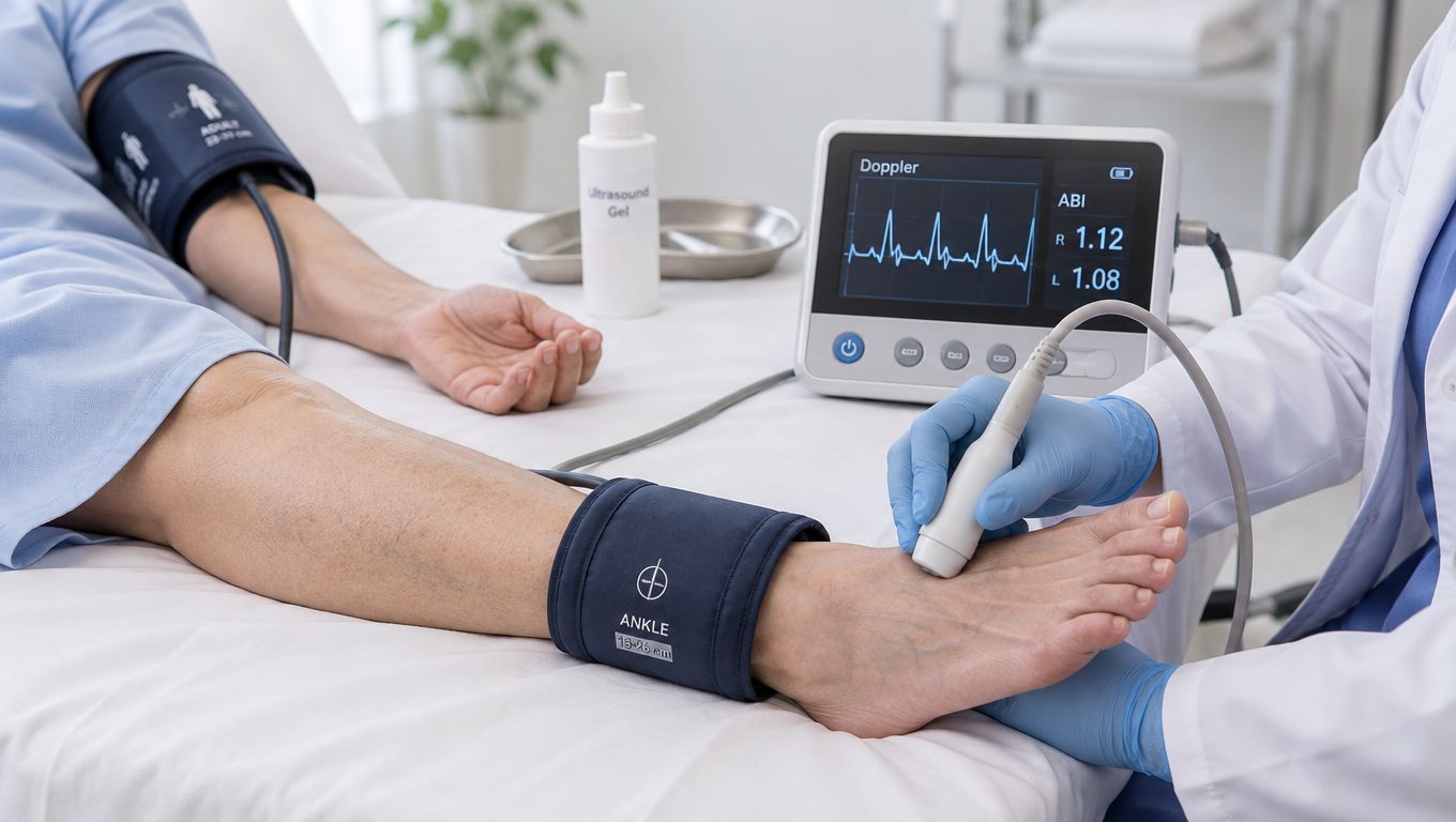
A busy Delaware executive can feel fit, exercise regularly, and still carry vascular disease that hasn't produced an obvious warning. Leg discomfort may be blamed on training or aging, while the underlying issue is reduced arterial flow. The ankle-brachial index, or ABI test, offers a straightforward way to compare blood pressure in the legs with blood pressure in the arms and place that finding in the context of whole-patient cardiovascular risk.
The test is noninvasive and uses blood-pressure cuffs with a Doppler device. Its value isn't limited to identifying circulation problems in the legs. A low or uninterpretable result can prompt a broader review of atherosclerosis risk, blood pressure, lipid patterns, diabetes, kidney health, exercise tolerance, and symptoms. The important clinical question isn't merely whether a resting number is “normal.” It's whether the result fits the person in front of the clinician.
Table of Contents
- Why Your Leg Vessels Are a Window Into Your Heart
- What the Ankle Brachial Index Actually Measures
- How a Resting ABI Test Is Performed
- Reading Your ABI Result the Right Way
- When an Exercise ABI Adds the Missing Answer
- What ABI Says About Cardiovascular and Longevity Risk
- Common Misinterpretations Worth Knowing
- Considering ABI in a Preventive Cardiology Visit
Why Your Leg Vessels Are a Window Into Your Heart
A 49-year-old executive may run marathons, maintain a demanding travel schedule, and feel entirely well while still having coronary calcium and silent peripheral atherosclerosis. That scenario captures a central challenge in preventive cardiology. Fitness can coexist with plaque, and the absence of leg pain doesn't prove that the leg arteries are free of disease.
The arteries supplying the legs are part of the same vascular system that supplies the heart and brain. Peripheral artery disease, or PAD, reflects arterial narrowing that can affect walking and limb health, but it also raises concern about atherosclerosis elsewhere. ABI helps clinicians look for that vascular signal with a brief comparison of arm and ankle pressures.
A simple test with systemic meaning
During a resting ABI assessment, a patient lies down while a clinician measures systolic pressure in both arms and at the arteries of both ankles. The calculation is a ratio, so a lower ankle pressure relative to the arm can indicate impaired arterial transmission to that leg. The method became standardized because it was reproducible, inexpensive, and practical across primary care, cardiology, and vascular medicine. The standardized calculation and classic diagnostic performance are described in this peer-reviewed ABI review.
Clinical perspective: A leg-vessel finding deserves a heart and brain risk review, not a narrow discussion about walking comfort alone.
That broader view matters for professionals in Lewes, Wilmington, Rehoboth, and nearby Pennsylvania or Maryland who may prioritize work capacity and long-term independence. Leg symptoms can be obvious, subtle, or absent. When symptoms do occur, resources explaining poor circulation treatment options can help readers understand the range of medical management, although the correct next step depends on clinical evaluation.
ABI is best understood as a risk lens rather than a verdict. Later sections will distinguish the four principal resting categories, explain why a high result can also be abnormal, and focus on exercise ABI, the diagnostic step many patient-facing explanations leave out.
What the Ankle Brachial Index Actually Measures
A patient may have no leg pain yet still show reduced arterial pressure at the ankle. The ankle-brachial index helps identify that pattern by comparing systolic pressure in each ankle with pressure in the arm. It functions like a pressure transmission check: healthy, compressible leg arteries usually carry arm-level pressure toward the feet, while narrowed or stiff vessels can alter the ratio.
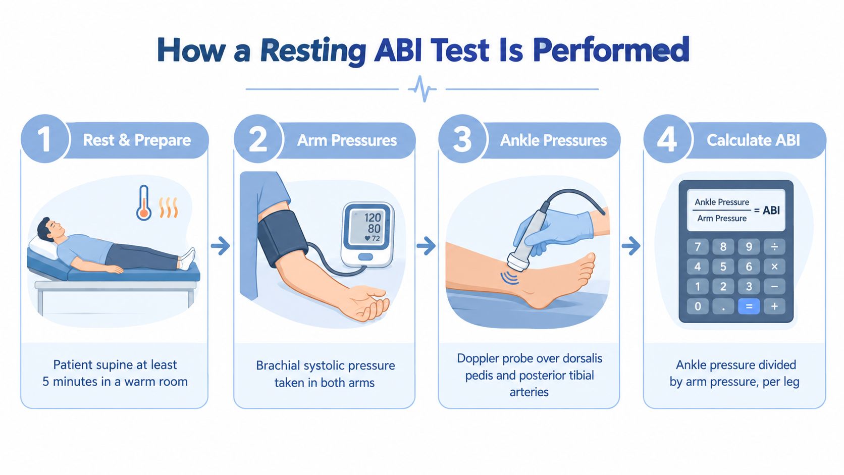
For each leg, the clinician selects the higher pressure recorded from the dorsalis pedis or posterior tibial artery and divides it by the higher brachial pressure. The right and left results are reported separately because disease may affect one leg more than the other. A Doppler probe detects blood flow when a standard blood-pressure method cannot reliably identify the arterial signal. Properly sized cuffs and stillness help capture resting circulation rather than a temporary response to movement.
A practical traffic-light interpretation
The result is easier to understand when viewed in clinical categories:
- Green, normal: 1.00 to 1.40, generally consistent with compressible arteries and no low-pressure pattern associated with PAD.
- Yellow, borderline: 0.91 to 0.99, a range that calls for symptoms and risk factors rather than automatic reassurance.
- Orange, abnormal: ≤0.90, consistent with PAD in the appropriate clinical setting.
- Red, more concerning: A substantially reduced value, especially below 0.70, indicates greater pressure loss and needs prompt medical context.
The AHA and ACC calculation uses the higher ankle pressure divided by the higher arm pressure for each leg. Its framework classifies ABI as abnormal at ≤0.90, borderline at 0.91 to 0.99, normal at 1.00 to 1.40, and noncompressible above 1.40. These categories are summarized in the current AHA and ACC PAD guideline.
A value above 1.40 does not mean circulation is excellent. Arterial walls may be too stiff to compress, often because of medial arterial calcification. Clinicians may then use another vascular test to clarify blood flow. ABI therefore offers more than a leg measurement. It can prompt a broader review of cardiovascular risk, even when walking symptoms are absent.
How a Resting ABI Test Is Performed
The resting procedure follows a defined sequence. The patient removes restrictive clothing around the arms and lower legs, lies flat, and avoids conversation or movement while the clinician obtains the measurements. The test doesn't involve needles, contrast dye, or radiation, and cuff inflation is usually brief, although it can feel snug.
The measurement sequence
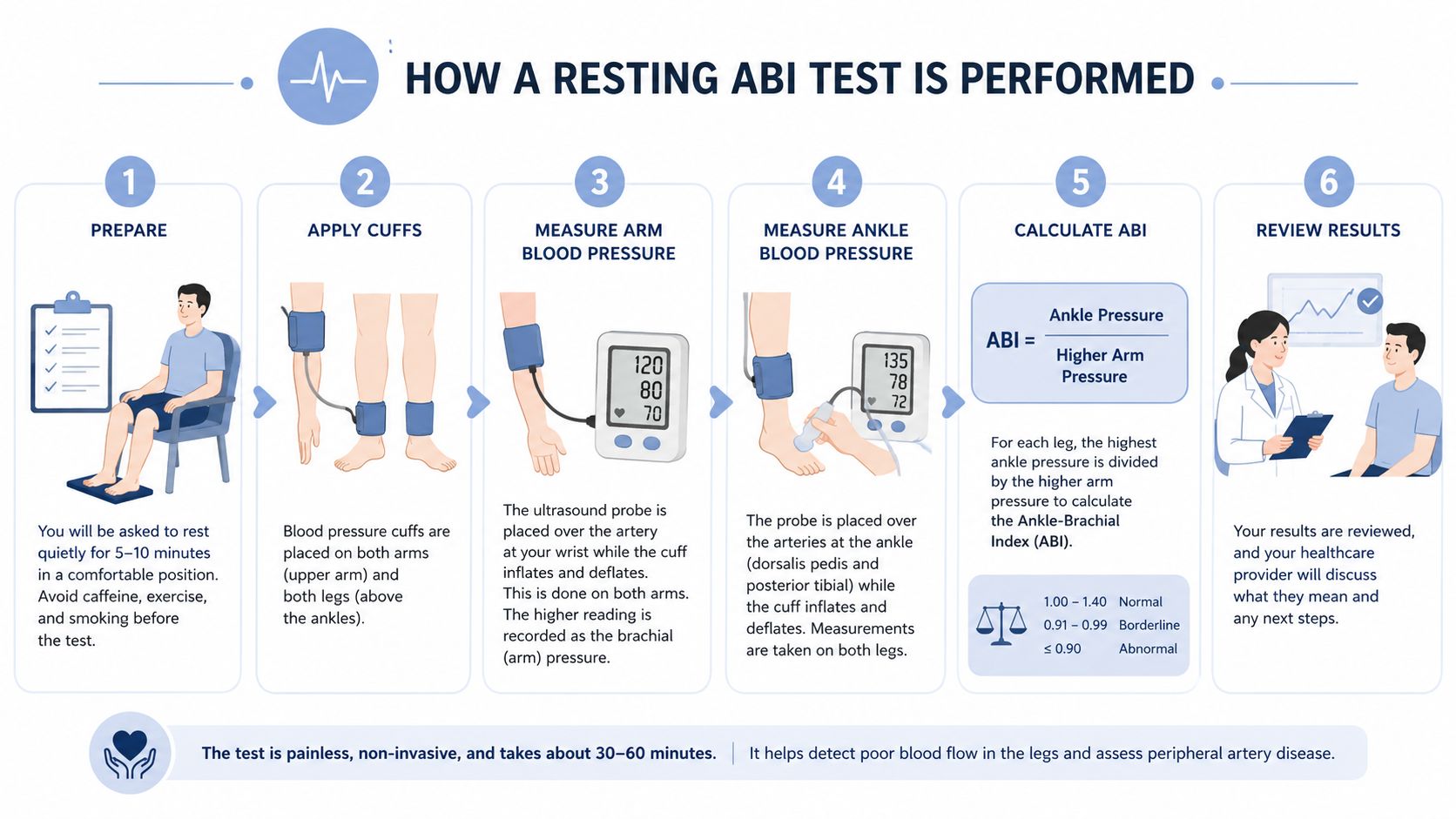
- Resting position: The patient rests supine for at least 5 minutes in a warm room. Warmth and quiet reduce avoidable changes in vascular tone.
- Arm pressures: A clinician measures systolic pressure in both arms with an appropriately sized cuff and handheld Doppler. The higher brachial pressure becomes the denominator for both leg calculations.
- Ankle pressures: A cuff sits just above the malleoli. The clinician records pressure at the dorsalis pedis and posterior tibial arteries in each leg.
- Leg-specific calculation: The higher ankle pressure from each leg is divided by the higher brachial pressure.
- Clinical comparison: The right and left ratios are reviewed independently. The lower ankle-brachial ratio usually receives the greatest attention because it reflects the more affected limb.
- Interpretation: The clinician combines the number with symptoms, pulses, risk factors, and examination findings.
Most patients can resume normal activity immediately. A normal experience feels much like repeated blood-pressure checks, with Doppler sounds replacing a stethoscope. The test can fit efficiently into an appointment, especially when a preventive cardiology team is already reviewing blood pressure, lipid markers, exercise history, and vascular symptoms.
For professionals evaluating a broader cardiovascular strategy, a preventive cardiologist near the patient's location can help place ABI findings alongside the rest of the risk profile.
Reading Your ABI Result the Right Way
An ABI is a ratio, not a standalone blood-pressure reading. A result of 0.80 means the measured ankle systolic pressure is lower than the higher arm pressure, expressed as that ratio. Each leg receives its own value, so the sides may differ. The lower result often deserves closer attention because it may identify the more affected limb.
Use the resting categories as a starting point, not a diagnosis by themselves. A borderline value carries more weight in someone with exertional calf discomfort, diabetes, kidney disease, smoking exposure, or known coronary disease than in a person without symptoms or other strong indicators.
| ABI Value | Interpretation | Clinical Meaning |
|---|---|---|
| 1.00 to 1.40 | Normal | The resting pressure relationship falls within the guideline-defined normal range, although symptoms may still require evaluation. |
| 0.91 to 0.99 | Borderline | The result is not clearly abnormal, but it may not explain exertional symptoms or resolve clinical concern. |
| 0.70 to 0.90 | Mild to moderate PAD pattern | Reduced ankle pressure supports PAD and calls for assessment of cardiovascular and limb risk. |
| Below 0.70 | More substantially reduced | The pressure deficit is greater and merits timely clinical evaluation. |
| Above 1.40 | Noncompressible | Arterial stiffness or calcification may prevent reliable cuff compression. |
In symptomatic patients, resting ABI sensitivity is reported as 69% to 79%, with specificity of 83% to 99%, compared with imaging-confirmed arterial stenosis. Those figures describe test performance, not an individual's certainty. Diabetes can reduce sensitivity because medial arterial calcification may produce falsely high ankle pressures.
A normal resting ABI answers a resting question. It may not show what happens when walking increases the legs' demand for blood flow.
That distinction matters clinically. Arterial symptoms with a normal or borderline resting result may call for exercise ABI testing, which can expose a pressure drop hidden while the patient is still.
When an Exercise ABI Adds the Missing Answer
Resting ABI measures circulation while the patient is lying still. Exercise ABI tests the same circulation under demand, when walking muscles require increased blood flow. That difference can expose flow-limiting disease that remains hidden at rest.
The AHA and ACC guidance recommends treadmill exercise ABI testing for patients with exertional, non-joint-related leg symptoms and a resting ABI above 0.90 through 1.40. The clinician generally records a resting baseline, has the patient walk on a treadmill or perform a defined active-standing protocol, and repeats ankle pressures during recovery, including at one and later recovery points when clinically appropriate.
Resting versus exercise testing
| Question | Resting ABI | Exercise ABI |
|---|---|---|
| Physiologic state | Lying quietly | After walking or exertion |
| Best use | Initial pressure comparison | Symptoms despite normal or borderline resting results |
| Diagnostic response | Low resting ratio | A post-exercise ABI drop of ≥20% or ankle pressure fall above 30 mmHg supports PAD |
| Clinical value | Detects resting pressure loss | Separates arterial claudication from pseudoclaudication and assesses walking limitation |
The exact threshold used in practice should follow the testing protocol and guideline interpretation. The AHA and ACC PAD guideline slide set identifies a post-exercise ABI decline of ≥20% or a post-exercise ankle systolic pressure fall above 30 mmHg as diagnostic in multiple expert sources.
Exercise testing is especially useful for symptomatic patients whose resting ABI is above 0.90, older adults with exertional leg symptoms, and higher-risk people whose resting values sit near the borderline cutoff. It also provides an objective view of walking performance in people with known PAD.
What ABI Says About Cardiovascular and Longevity Risk
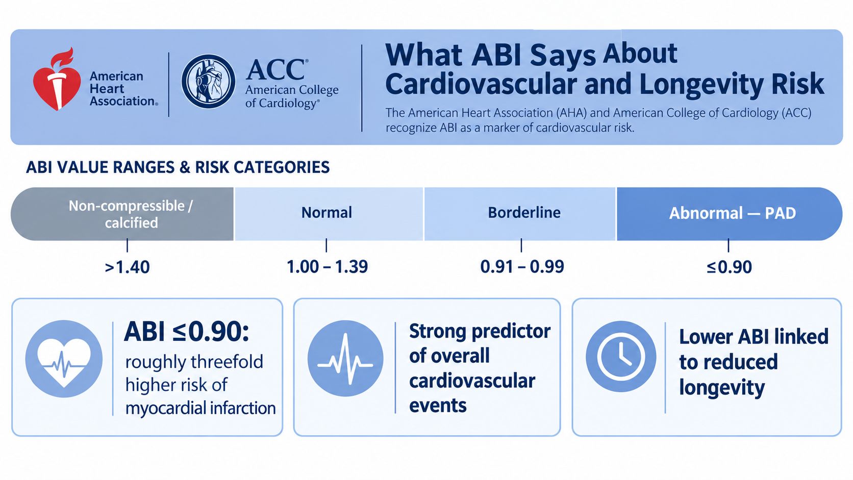
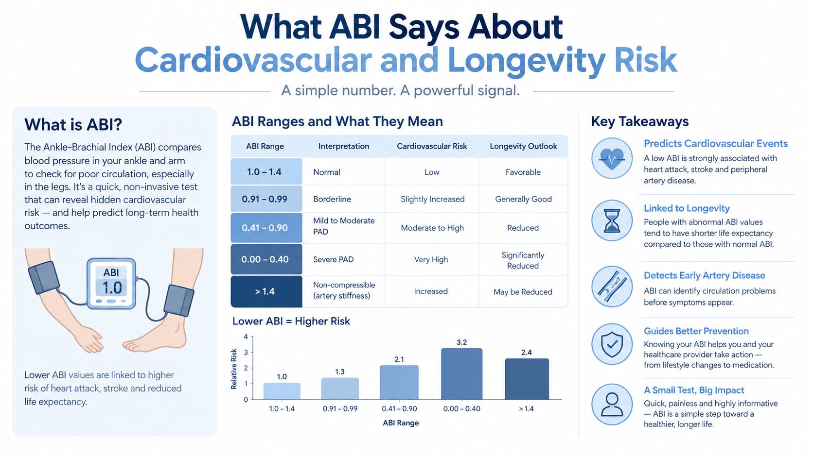
ABI findings connect the leg arteries to the broader cardiovascular system. A reduced result indicates more than a local circulation problem. It can signal atherosclerotic burden that deserves review across the coronary, carotid, renal, and peripheral vascular beds.
The AHA and ACC framework treats an abnormal ABI as a marker of cardiovascular risk. Clinical summaries report that an ABI under 0.90 has about 95% sensitivity and 100% specificity versus angiography in some clinical reviews, while other summaries place sensitivity between 80% and 95% and specificity around 95% to 100%. Those figures describe diagnostic performance for PAD, not a personal prediction of an individual's future.
Low and high results both matter
A low ABI can reclassify a person whose traditional risk estimate appears intermediate, particularly when symptoms or other evidence point toward systemic atherosclerosis. The clinical response may include a closer review of preventive treatment, exercise capacity, smoking exposure, glucose regulation, lipids, blood pressure, and symptoms. It doesn't guarantee an outcome or determine a treatment plan by itself.
A high ABI above 1.40 raises a different concern. Noncompressible arteries can reflect medial arterial calcification, which is more likely in people with diabetes, chronic kidney disease, or advanced age. A deceptively high ratio may therefore require toe-brachial index testing or another method rather than reassurance.
For readers building a broader framework for healthy aging and risk reduction, this longevity guide for adults over 40 offers general context. ABI should sit within a physician-led prevention plan, alongside appropriate imaging and laboratory evaluation. A related overview of advanced cardiovascular imaging explains how vascular findings can be integrated with other forms of risk assessment.
Common Misinterpretations Worth Knowing
ABI is powerful because it's simple. That simplicity also creates traps when people treat a single number as a complete diagnosis.
Trap one, a normal resting result ends the evaluation
A resting ABI above 0.90 can miss disease that becomes apparent only during exertion. The problem is particularly relevant when a patient reports reproducible calf, thigh, or buttock discomfort during walking that improves with rest, but the resting measurement doesn't show a clear abnormality.
The Cleveland Clinic ABI explanation notes the commonly used normal and abnormal ranges, while guideline-style sources support post-exercise testing when clinical suspicion persists. In selected screen-detected older populations, ABI sensitivity versus magnetic resonance angiography has been reported as low as 7% to 34%, and another review describes a false-negative rate of about 20% for the 0.90 cutoff. These figures show why symptoms and pre-test probability matter.
Trap two, a high ABI means healthier arteries
Arteries above 1.40 may be noncompressible. A cuff can't reliably collapse a heavily calcified vessel, so the resulting pressure can look falsely high. Diabetes, chronic kidney disease, and older age increase the importance of this limitation, and clinicians may consider toe pressures, toe-brachial index, segmental pressures, pulse-volume recordings, or imaging.
Trap three, everyone should receive broad screening
The evidence doesn't support treating ABI as an automatic population-wide test for every asymptomatic adult. The USPSTF has found evidence insufficient for screening asymptomatic adults overall, while more targeted guidance considers testing when risk factors, symptoms, known atherosclerosis, or a management decision make the result clinically meaningful.
Interpretation rule: When the number and the symptoms disagree, the disagreement is the reason to investigate further.
Considering ABI in a Preventive Cardiology Visit
ABI belongs in preventive cardiology when it can answer a meaningful vascular question. It isn't reserved for people already seeing a vascular surgeon, and it shouldn't be ordered without a plan for interpreting the result. The decision usually begins with age, symptoms, examination findings, smoking history, diabetes, kidney disease, blood pressure, lipid markers, and known atherosclerosis.
People who may merit a clinician discussion include:
- Older adults: Adults over 65 may warrant consideration based on their overall risk profile.
- Risk-factor burden: Adults over 50 with cardiovascular risk factors may benefit when the result could change management.
- Exertional symptoms: Leg pain, heaviness, cramping, or fatigue during walking deserves evaluation, especially when it improves with rest.
- Tissue concerns: Nonhealing wounds or foot changes require medical assessment rather than self-monitoring.
- Established disease: Known coronary, carotid, or renal atherosclerosis increases the relevance of a peripheral vascular assessment.
- Selected high-risk patients: Smokers or people with diabetes and additional risk markers may need a more targeted approach.
What a focused visit can include
A preventive cardiology visit may start with a focused vascular history and examination, followed by a supine resting ABI with Doppler. If the resting result is borderline, unexpectedly normal despite symptoms, or above the compressibility range, the clinician can consider exercise ABI, toe-brachial index, segmental pressure testing, pulse-volume recordings, or imaging.
At Hosmane Longevity Center, the physician-led preventive evaluation can integrate ABI-related assessment with coronary calcium scoring, carotid evaluation, lipid analysis, blood-pressure profiling, and other advanced cardiac testing when clinically appropriate. Testing is available to people who aren't executive health program members, subject to the practice's consultation and care model.
The purpose is not to collect more measurements. It's to decide which findings matter, which require confirmation, and which preventive actions deserve priority. A person with a family history of premature disease can also review risk context through this discussion of family history and heart disease screening.
Hosmane Longevity Center offers physician-led preventive cardiology consultations and advanced vascular assessment, including resting and exercise ABI evaluation when clinically appropriate. Adults in Delaware and nearby Pennsylvania or Maryland can visit Hosmane Longevity Center to discuss whether ABI belongs in a personalized cardiovascular and longevity assessment.