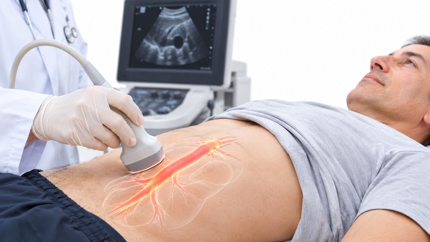
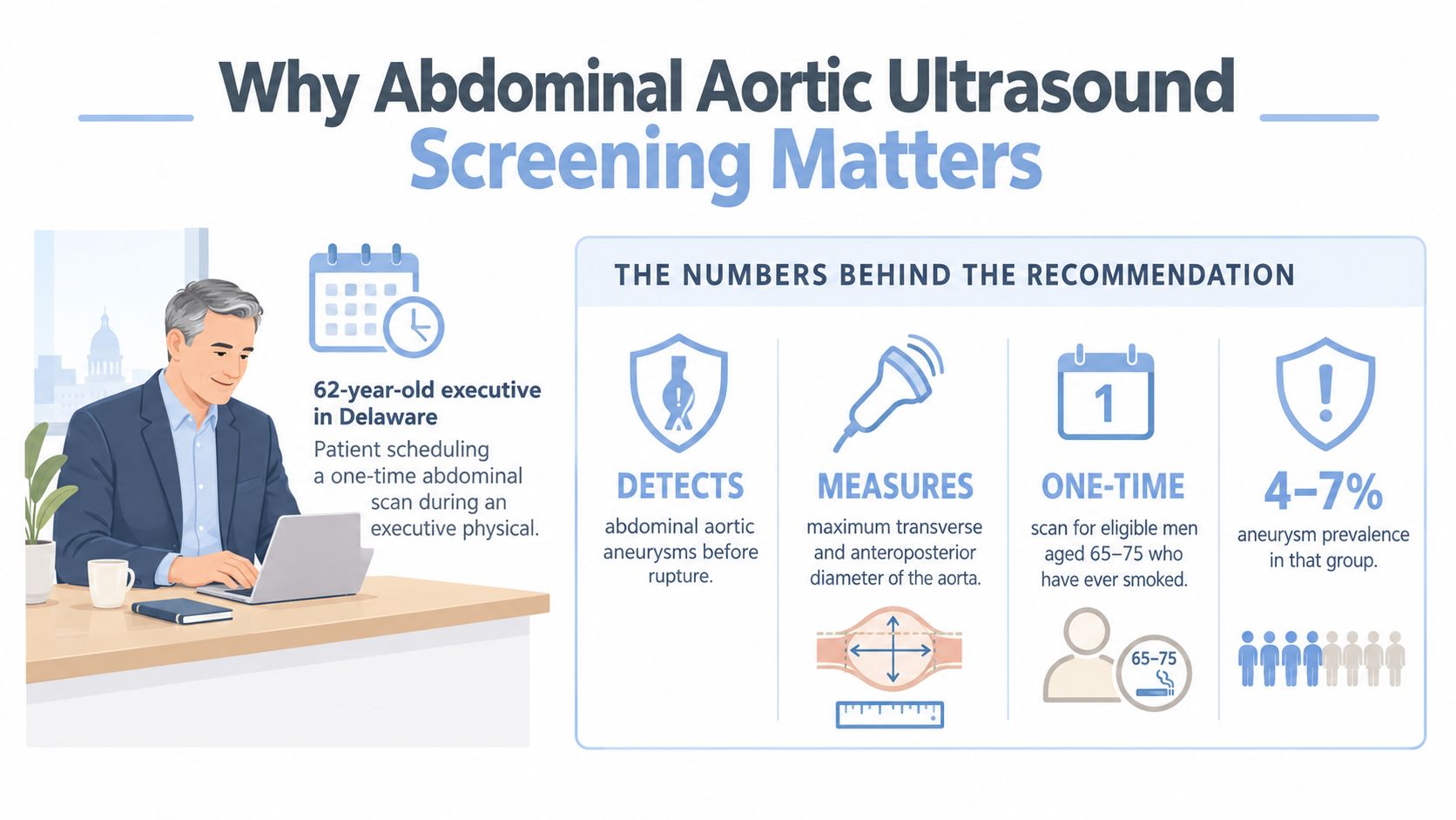
A 62-year-old executive in Delaware has a packed calendar, a family history of vascular disease, and no symptoms that suggest an aortic problem. During an executive physical, the question is whether a one-time abdominal scan adds useful information or merely creates another result to interpret.
That question deserves a measured answer. Abdominal aortic ultrasound screening can identify enlargement before rupture, but the scan isn't a verdict and it isn't appropriate for everyone. Its value comes from the number it measures, the patient's risk profile, and the follow-up plan attached to the result.
Table of Contents
- Why Abdominal Aortic Ultrasound Screening Matters
- What an Abdominal Aortic Aneurysm Actually Is
- Who Should Be Screened Under Current Guidelines
- How the Ultrasound Test Is Performed
- Reading the Result and What the Numbers Mean
- Why More Screening Is Not Always Better
- Next Steps When a Scan Shows an Aneurysm
- Adding Ultrasound to a Preventive Cardiology Plan
Why Abdominal Aortic Ultrasound Screening Matters
The abdominal aorta is the large artery that carries blood from the heart through the abdomen toward the lower body. An aneurysm develops when a section of its wall enlarges. Because an abdominal aortic aneurysm, or AAA, can remain silent, a person may feel completely well while the aorta gradually changes.
Ultrasound measures the maximum diameter of the abdominal aorta, particularly the infrarenal segment below the kidney arteries. That measurement is a practical surrogate for rupture risk because larger aneurysms generally require closer surveillance and may eventually require vascular specialist assessment.
The landmark United Kingdom Multicentre Aneurysm Screening Study randomized 67,770 men aged 65 to 74 to an invitation for one-time ultrasound or to a control group. After 10 years, AAA-related deaths occurred in 155 men in the invited group, an absolute risk of 0.46%, compared with 296 deaths, or 0.87%, in the control group. The relative reduction in AAA-related mortality was 48%, with a hazard ratio of 0.52 and a 95% confidence interval of 0.43 to 0.63, as reported in the MASS trial review.
The result needs careful interpretation. Total mortality was approximately 30% in both groups, because AAA accounted for only a small proportion of deaths in this age range. Ultrasound therefore has a specific purpose, preventing rupture-related death through detection and management, rather than proving that screening extends overall lifespan.
Practical rule: The scan produces a measurement. The measurement produces a risk category. The risk category determines whether the next step is reassurance, prevention, surveillance, or referral.
For a professional in Lewes, Wilmington, Rehoboth, or nearby Pennsylvania or Maryland, that distinction matters. A normal result may close the AAA question for a long time, while an enlarged result should enter a longitudinal vascular-risk record rather than sit alone in an executive health file.
What an Abdominal Aortic Aneurysm Actually Is
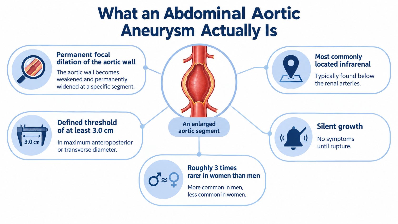
An abdominal aortic aneurysm is a permanent, focal enlargement of part of the abdominal aortic wall. It most often affects the infrarenal aorta. In clinical practice, 3.0 cm or greater is commonly used as the lower boundary for an AAA, according to the 2022 ACC/AHA aortic disease guideline.
The definition is based on measurement, not on whether a person has pain. A clinician generally wants the maximum anteroposterior or transverse diameter, using a consistent technique. A mildly enlarged aorta may produce no sensation at all, while a symptomatic aneurysm can require urgent evaluation. Symptoms aren't a reliable screening tool.
Why diameter drives decisions
A larger radius increases the mechanical tension carried by the arterial wall. This relationship, often explained through Laplace's law, helps clarify why rupture risk doesn't rise in a simple, linear fashion as an aneurysm expands. The size of the aorta, its shape, growth rate, symptoms, and the patient's operative risk all influence management.
Several terms are often confused:
- True aneurysm: Enlargement involving the layers of the arterial wall.
- Ectasia: Mild enlargement that doesn't meet the usual AAA definition.
- Pseudoaneurysm: A contained leak or disruption outside the normal full wall structure.
- Dissection: A tear that allows blood to track between layers of the aortic wall.
Smoking-related oxidative injury, inflammation, and degradation of structural proteins such as elastin and collagen contribute to weakening of the aortic wall. These mechanisms explain why smoking history remains clinically important, but they don't allow a clinician to predict an individual result from one risk factor alone.
An AAA is best understood as a structural finding with a measurable growth rate. Ultrasound is useful because it can document that structure repeatedly without radiation. A report that states only “mild enlargement” isn't enough for a durable care plan. The exact diameter, image quality, location, and comparison with prior measurements are more clinically useful.
Who Should Be Screened Under Current Guidelines
Screening recommendations aren't interchangeable. The U.S. Preventive Services Task Force focuses on the balance between expected benefit and potential harm in asymptomatic adults, while the American College of Cardiology and American Heart Association framework also gives attention to vascular disease, family history, and previous aortic conditions.
The USPSTF recommends a one-time ultrasound for asymptomatic men aged 65 to 75 who have ever smoked, a Grade B recommendation. In the relevant evidence base, “ever smoked” means at least 100 cigarettes over a lifetime. For men in the same age range who never smoked, screening is selective rather than routine. The task force recommends against routine screening in women who have never smoked and finds insufficient evidence for or against screening women aged 65 to 75 who have ever smoked, as outlined in the current USPSTF recommendation.
The ACC/AHA approach can support individualized assessment for people with a first-degree relative with AAA, peripheral arterial disease, or a history of aortic repair. Such situations don't mean that every person needs immediate imaging. They mean the decision should account for the person's vascular history rather than relying only on age and smoking status.
Comparing the frameworks
| Population | USPSTF Recommendation | ACC/AHA Recommendation |
|---|---|---|
| Men aged 65 to 75 who have ever smoked | One-time ultrasound, Grade B | Screening is supported within a broader vascular-risk assessment |
| Men aged 65 to 75 who never smoked | Selective screening, Grade C | Individualized assessment based on vascular disease and family history |
| Women aged 65 to 75 who have never smoked | Routine screening isn't recommended | Individualized assessment may be considered when risk is clinically significant |
| Women aged 65 to 75 who have ever smoked | Evidence is insufficient for a universal recommendation | Consider risk factors, family history, and vascular disease |
| Adults with a first-degree relative with AAA, peripheral arterial disease, or prior aortic repair | Not fully defined by the basic population recommendation | A more individualized screening discussion may be appropriate |
Smoking history is the dominant modifiable driver, followed by age, sex, family history, hypertension, and dyslipidemia. A 62-year-old former smoker with peripheral arterial disease may have a different rationale for imaging than a 62-year-old never-smoker without vascular risk.
A family history of cardiovascular disease also deserves a broader review, as discussed in this guide to family history and heart disease screening. A preventive cardiologist may reasonably offer screening outside the strict USPSTF population when the expected information could change surveillance or risk management. That decision should be documented as individualized care, not presented as a universal screening rule.
How the Ultrasound Test Is Performed
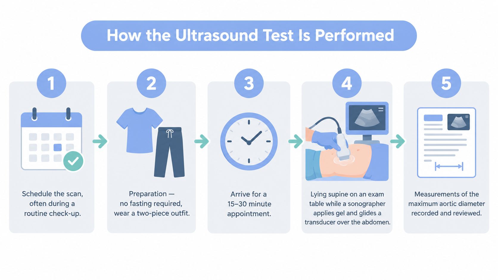
The examination is usually straightforward and noninvasive. Scheduling staff confirm the clinical reason for the scan, relevant risk factors, and whether earlier imaging is available for comparison.
Most abdominal aortic ultrasound examinations don't require fasting. A two-piece outfit makes it easier to expose the abdomen while preserving comfort and privacy. The appointment commonly takes 15 to 30 minutes, depending on anatomy, image quality, and the number of views required.
The scanning sequence
The patient lies on a table in a supine position. The sonographer applies gel and moves a curvilinear probe across the abdomen, generally from the upper abdominal region toward the umbilicus. B-mode grayscale imaging creates the primary views, including transverse and longitudinal images of the aorta.
Color Doppler may help confirm blood flow and distinguish moving blood from slow flow or thrombus along the vessel wall. This distinction is critical because an aneurysm can contain mural thrombus, and the outer wall measurement must not be confused with the open channel of flowing blood.
The sonographer then records the maximum anterior-posterior diameter, usually at the infrarenal segment. The report should identify the measurement technique and whether the study was technically complete. Obesity, bowel gas, or other factors can limit visualization, so a technically limited examination shouldn't automatically be treated as a definitive negative result.
What happens after the scan
A qualified reader, such as a radiologist or vascular laboratory physician, reviews the images and measurements. The structured report should describe the aortic diameter, the segment examined, image quality, and any relevant associated findings.
The referring clinician then interprets the result in context. Advanced cardiovascular imaging can be most useful when the report reaches the same physician who is tracking blood pressure, lipid markers, smoking exposure, and other vascular information. A broader overview of how imaging can fit into prevention appears in this guide to advanced cardiovascular imaging.
Reading the Result and What the Numbers Mean
A report becomes clinically useful when its number leads to a defined action. An aorta below the usual AAA threshold is generally treated differently from an aorta that requires serial surveillance. The Society for Vascular Surgery surveillance guidance and the ACC/AHA guideline use diameter-based intervals, although local pathways can differ.
For orientation, an abdominal aorta below 3.0 cm in men and below 2.7 cm in women is generally considered within the usual non-aneurysmal range. A diameter of 3.0 to 3.9 cm typically leads to longer-interval surveillance, while larger measurements require progressively closer review.
| Diameter in men | Diameter in women | Surveillance interval | Clinical action |
|---|---|---|---|
| Below 3.0 cm | Below 2.7 cm | No routine repeat solely for AAA in many patients | Document the result and address vascular risk |
| 3.0 to 3.9 cm | 3.0 to 3.9 cm | Often approximately every 3 years under SVS guidance | Establish surveillance and risk-reduction plan |
| 4.0 to 4.9 cm | 4.0 to 4.4 cm | Approximately annually, depending on protocol | Continue structured surveillance and consider vascular input |
| 5.0 to 5.4 cm | 4.5 to 5.4 cm | Approximately every 6 months, or more intensively under some pathways | Vascular surgery consultation and repair assessment |
| At least 5.5 cm | At least 5.0 cm in women | Specialist-directed | Consider elective repair based on anatomy, symptoms, growth, and operative risk |
The United Kingdom pathway follows a more intensive schedule, with annual follow-up for 3.0 to 4.4 cm, quarterly follow-up for 4.5 to 5.4 cm, and specialist referral at 5.5 cm or larger, according to its 10-year effectiveness review. These differences don't represent a contradiction. They show why the governing protocol and specialist judgment must accompany the measurement.
What else belongs in the report
Diameter is central, but it isn't the only feature that matters. The report may comment on:
- Shape, such as fusiform or saccular enlargement.
- Iliac involvement, which can affect future planning.
- Mural thrombus, which may line part of the aneurysm.
- Growth compared with prior studies, interpreted alongside measurement variability.
- Image quality, including whether bowel gas or body habitus limited the examination.
In men, 5.5 cm is a commonly used elective repair threshold, while in women 5.0 cm is commonly used. Symptoms, anatomy, operative risk, and unusually rapid expansion can change the timing. A measurement should therefore trigger a conversation, not an automatic operation.
Why More Screening Is Not Always Better
A common executive-health instinct is to repeat every test annually. That approach can be sensible for some biomarkers, but it isn't automatically appropriate for abdominal aortic ultrasound screening.
The strongest evidence for population screening centers on men aged 65 to 75 who have ever smoked. The USPSTF gives selective consideration to men in that age band who never smoked, while the evidence remains insufficient for routine screening in women who have never smoked. The USPSTF evidence summary also evaluates repeat screening, treatment of small AAAs, and potential harms.
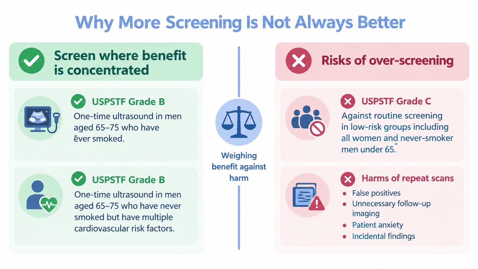
The cost of indiscriminate repetition
Low-risk screening can produce borderline measurements, uncertain findings, and anxiety without a demonstrated mortality benefit. Follow-up imaging may expose patients to additional procedures or, in some cases, CT imaging when ultrasound cannot answer the question clearly.
A stable aorta measuring 2.8 cm doesn't usually justify an annual rescan solely because the patient has access to an executive health program. That time and attention may be more valuable when directed toward blood-pressure control, lipid management, physical activity, or smoking cessation.
More data isn't automatically better prevention. The right interval is the one that can detect meaningful change without creating a cycle of low-value testing.
The principle isn't anti-ultrasound. It is risk-stratified use. A scan should be ordered when the patient's risk and the expected clinical decision justify it, then repeated according to the measured diameter, growth pattern, technical quality, and governing protocol.
Next Steps When a Scan Shows an Aneurysm
A positive scan usually leads first to confirmation and classification, not immediate surgery. The clinician verifies the maximum diameter, reviews prior imaging, checks whether the study was technically adequate, and determines whether the finding is a true aneurysm or another form of aortic abnormality.
The referral pathway
A vascular surgery consultation becomes increasingly important as the diameter grows, the growth rate accelerates, symptoms appear, or the anatomy is complex. The referral packet should include:
- The complete ultrasound report and representative images.
- Prior aortic measurements, if available.
- Smoking history and current medications.
- Blood-pressure readings and lipid results.
- Relevant cardiovascular, peripheral arterial, and family history.
- Kidney function and other information needed for contrast-based imaging.
CT angiography may be used for operative planning when repair is under consideration. A cardiopulmonary assessment helps determine procedural risk, while a preventive review identifies modifiable factors that can be addressed before any elective intervention.
Repair is generally considered around 5.5 cm in men or 5.0 cm in women, with earlier action possible for symptoms, anatomy, or rapid enlargement. Expansion of more than 0.5 cm in 6 months is a concerning pattern that can accelerate specialist decision-making. These thresholds are not promises or automatic orders. The vascular surgeon weighs them against anatomy, operative risk, symptoms, and the patient's preferences.
Medical management continues
A small or moderate aneurysm still belongs in an active prevention plan. Smoking cessation is a central intervention. Blood-pressure control, lipid management, physical activity appropriate to the patient's condition, and antiplatelet decisions when otherwise indicated all require clinician oversight.
A preventive cardiologist can coordinate the imaging interval, communicate the significance of the measurement, review the broader vascular profile, and help optimize the patient before elective repair if repair becomes appropriate. The goal is not to make the aneurysm disappear through lifestyle changes. The goal is to reduce avoidable vascular risk while the aorta is monitored properly.
Adding Ultrasound to a Preventive Cardiology Plan
An abdominal aortic ultrasound is one data point in a broader vascular-risk strategy. It answers a focused question about aortic structure. It doesn't replace assessment of coronary plaque, blood pressure, lipid particles, glucose regulation, fitness, sleep, or family history.
For a high-demand professional, the most useful plan connects findings across time. A clinician may place the aortic measurement alongside:
- Coronary artery calcium scoring.
- Carotid intima-media thickness.
- Lipoprotein(a) and apolipoprotein B.
- Blood-pressure trajectory, including ambulatory monitoring when appropriate.
- Metabolic markers and exercise capacity.
- Sleep quality, stress load, nutrition, and alcohol intake.
The exact combination should follow the patient's risk profile rather than a fixed testing bundle. A finding of aortic enlargement might increase attention to smoking cessation and blood-pressure control. A high lipid burden may influence the discussion of statin intensity or other preventive treatment. Neither decision should be made from the ultrasound alone.
A longitudinal record also prevents a common error, treating every new measurement as if it were the first. Consistent technique, comparable imaging, and clear documentation help clinicians distinguish genuine growth from ordinary measurement variation.
Hosmane Longevity Center offers abdominal aortic ultrasound as an in-house cardiovascular imaging service, with findings reviewed as part of preventive cardiology and executive health planning. Testing is available to people who aren't program members as well, and a consultation can help determine whether a scan, surveillance plan, or broader vascular assessment is appropriate.
Readers in Delaware and nearby Pennsylvania or Maryland can visit Hosmane Longevity Center to discuss abdominal aortic ultrasound screening, advanced cardiac testing, and a coordinated preventive cardiology evaluation. The consultation can connect the aortic measurement with blood pressure, lipid markers, imaging findings, and practical next steps for a demanding schedule.